Home
/ Compact Bone Diagram Lacunae - 6 3 Bone Structure Anatomy Physiology _ (b) in this micrograph of the osteon, you can clearly see the concentric lamellae and central canals.
Compact Bone Diagram Lacunae - 6 3 Bone Structure Anatomy Physiology _ (b) in this micrograph of the osteon, you can clearly see the concentric lamellae and central canals.
Compact Bone Diagram Lacunae - 6 3 Bone Structure Anatomy Physiology _ (b) in this micrograph of the osteon, you can clearly see the concentric lamellae and central canals.. Haversian canals i (sometimes canals of havers ) are a series of microscopic tubes in the outermost region of bone called cortical bone. Jan 05, 2013 · lacunae are small cavities or chambers located between one lamella and the next. Diagram of a typical long bone showing both compact (cortical) and cancellous (spongy) bone. The lacunae and their osteocytes are present in trabeculae matrix on the bone with the bone marrow. Like compact bone, spongy bone, also known as cancellous bone, contains osteocytes housed in lacunae, but they are not arranged in concentric circles.
Jan 05, 2013 · lacunae are small cavities or chambers located between one lamella and the next. Sep 25, 2019 · the spongy bone is made up of cells, known as osteocytes which lie in small cavities called lacunae. (b) in this micrograph of the osteon, you can clearly see the concentric lamellae and central canals. Like compact bone, spongy bone, also known as cancellous bone, contains osteocytes housed in lacunae, but they are not arranged in concentric circles. Blood vessels travel from the harder compact bone toward the spongy bone, by supply the materials which are necessary for the production of blood.
Compact Bone And Spongy Bone Anatomy Flashcards Quizlet from o.quizlet.com (b) in this micrograph of the osteon, you can clearly see the concentric lamellae and central canals. The lacunae and their osteocytes are present in trabeculae matrix on the bone with the bone marrow. (b) in this micrograph of the osteon, you can clearly see the concentric lamellae and central canals. There are pores and spaces even in compact bone. Blood vessels travel from the harder compact bone toward the spongy bone, by supply the materials which are necessary for the production of blood. Diagram of a typical long bone showing both compact (cortical) and cancellous (spongy) bone. Like compact bone, spongy bone, also known as cancellous bone, contains osteocytes housed in lacunae, but they are not arranged in concentric circles. Consists of a central canal (haversian canal) surrounded by lamellar bone matrix within which osteocytes reside.
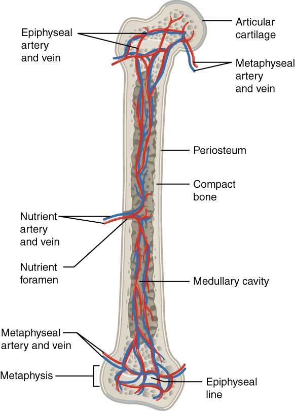
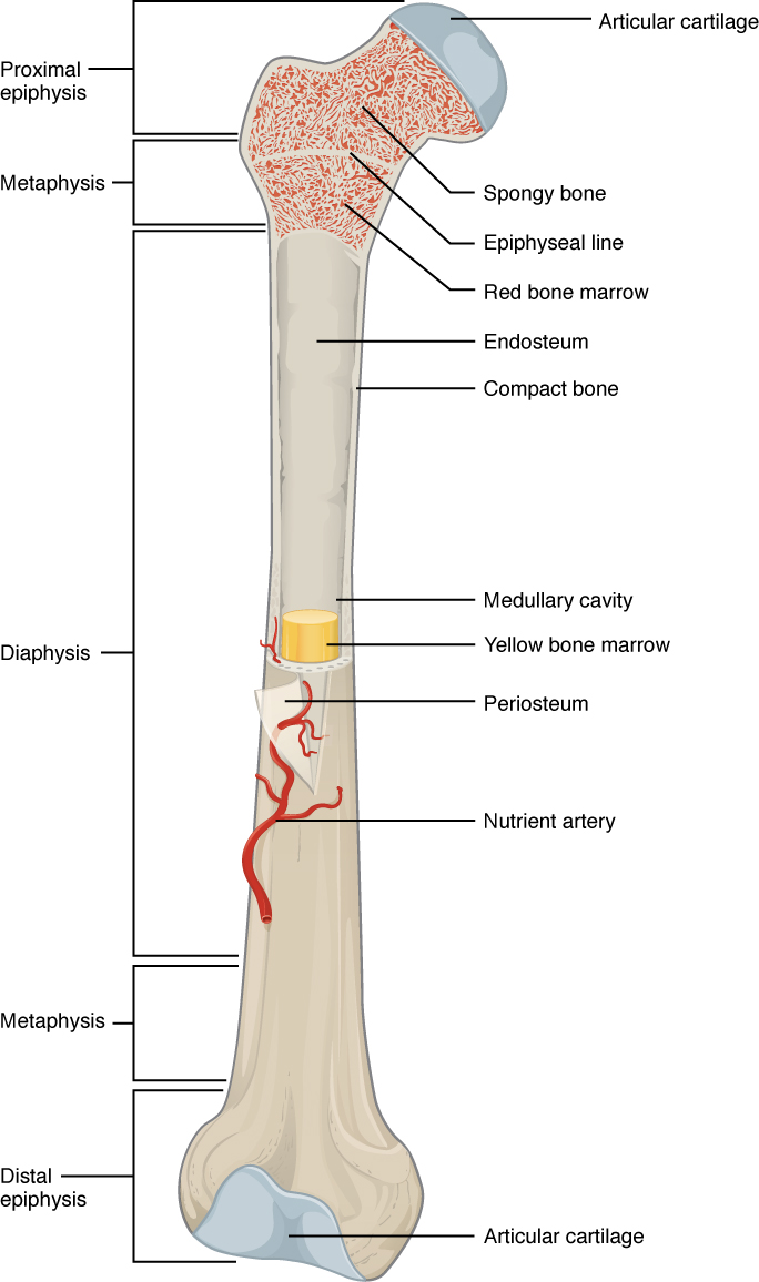
Diagram of a typical long bone showing both cortical (compact) and cancellous (spongy) bone.
Consists of a central canal (haversian canal) surrounded by lamellar bone matrix within which osteocytes reside. Like compact bone, spongy bone, also known as cancellous bone, contains osteocytes housed in lacunae, but they are not arranged in concentric circles. The lacunae and their osteocytes are present in trabeculae matrix on the bone with the bone marrow. (b) in this micrograph of the osteon, you can clearly see the concentric lamellae and central canals. Blood vessels travel from the harder compact bone toward the spongy bone, by supply the materials which are necessary for the production of blood. Sep 25, 2019 · the spongy bone is made up of cells, known as osteocytes which lie in small cavities called lacunae. Ʒ ən / (named for clopton havers ) is the fundamental functional unit of much compact bone. Haversian canals i (sometimes canals of havers ) are a series of microscopic tubes in the outermost region of bone called cortical bone. (b) in this micrograph of the osteon, you can clearly see the concentric lamellae and central canals. They have long extensions that project into the canaliculi. Jan 05, 2013 · lacunae are small cavities or chambers located between one lamella and the next. (the dark purple structures in the diagram above are the lacunae.) the osteocytes or mature bone cells are located in the lacunae. Diagram of a typical long bone showing both cortical (compact) and cancellous (spongy) bone.
Blood vessels travel from the harder compact bone toward the spongy bone, by supply the materials which are necessary for the production of blood. They have long extensions that project into the canaliculi. Sep 25, 2019 · the spongy bone is made up of cells, known as osteocytes which lie in small cavities called lacunae. Like compact bone, spongy bone, also known as cancellous bone, contains osteocytes housed in lacunae, but they are not arranged in concentric circles. The lacunae and their osteocytes are present in trabeculae matrix on the bone with the bone marrow.
What Is The Structure And Function Of The Compact Bone Socratic from opentextbc.ca (b) in this micrograph of the osteon, you can clearly see the concentric lamellae and central canals. (the dark purple structures in the diagram above are the lacunae.) the osteocytes or mature bone cells are located in the lacunae. Ʒ ən / (named for clopton havers ) is the fundamental functional unit of much compact bone. Sep 25, 2019 · the spongy bone is made up of cells, known as osteocytes which lie in small cavities called lacunae. Diagram of a typical long bone showing both compact (cortical) and cancellous (spongy) bone. There are pores and spaces even in compact bone. (b) in this micrograph of the osteon, you can clearly see the concentric lamellae and central canals. Jan 05, 2013 · lacunae are small cavities or chambers located between one lamella and the next.
They have long extensions that project into the canaliculi.
There are pores and spaces even in compact bone. They have long extensions that project into the canaliculi. The lacunae and their osteocytes are present in trabeculae matrix on the bone with the bone marrow. Haversian canals i (sometimes canals of havers ) are a series of microscopic tubes in the outermost region of bone called cortical bone. Jan 05, 2013 · lacunae are small cavities or chambers located between one lamella and the next. Blood vessels travel from the harder compact bone toward the spongy bone, by supply the materials which are necessary for the production of blood. Like compact bone, spongy bone, also known as cancellous bone, contains osteocytes housed in lacunae, but they are not arranged in concentric circles. Sep 25, 2019 · the spongy bone is made up of cells, known as osteocytes which lie in small cavities called lacunae. (b) in this micrograph of the osteon, you can clearly see the concentric lamellae and central canals. Like compact bone, spongy bone, also known as cancellous bone, contains osteocytes housed in lacunae, but they are not arranged in concentric circles. (b) in this micrograph of the osteon, you can clearly see the concentric lamellae and central canals. Consists of a central canal (haversian canal) surrounded by lamellar bone matrix within which osteocytes reside. Ʒ ən / (named for clopton havers ) is the fundamental functional unit of much compact bone.
Blood vessels travel from the harder compact bone toward the spongy bone, by supply the materials which are necessary for the production of blood. (the dark purple structures in the diagram above are the lacunae.) the osteocytes or mature bone cells are located in the lacunae. Like compact bone, spongy bone, also known as cancellous bone, contains osteocytes housed in lacunae, but they are not arranged in concentric circles. (b) in this micrograph of the osteon, you can clearly see the concentric lamellae and central canals. They have long extensions that project into the canaliculi.
Bones Fundamentals Of Anatomy For Physicians Lecturio from d3uigcfkiiww0g.cloudfront.net Like compact bone, spongy bone, also known as cancellous bone, contains osteocytes housed in lacunae, but they are not arranged in concentric circles. Diagram of a typical long bone showing both cortical (compact) and cancellous (spongy) bone. Blood vessels travel from the harder compact bone toward the spongy bone, by supply the materials which are necessary for the production of blood. (the dark purple structures in the diagram above are the lacunae.) the osteocytes or mature bone cells are located in the lacunae. Diagram of a typical long bone showing both compact (cortical) and cancellous (spongy) bone. (b) in this micrograph of the osteon, you can clearly see the concentric lamellae and central canals. There are pores and spaces even in compact bone. Ʒ ən / (named for clopton havers ) is the fundamental functional unit of much compact bone.
They have long extensions that project into the canaliculi.
Jan 05, 2013 · lacunae are small cavities or chambers located between one lamella and the next. Haversian canals i (sometimes canals of havers ) are a series of microscopic tubes in the outermost region of bone called cortical bone. Sep 25, 2019 · the spongy bone is made up of cells, known as osteocytes which lie in small cavities called lacunae. (the dark purple structures in the diagram above are the lacunae.) the osteocytes or mature bone cells are located in the lacunae. They have long extensions that project into the canaliculi. Ʒ ən / (named for clopton havers ) is the fundamental functional unit of much compact bone. Diagram of a typical long bone showing both cortical (compact) and cancellous (spongy) bone. There are pores and spaces even in compact bone. Diagram of a typical long bone showing both compact (cortical) and cancellous (spongy) bone. Like compact bone, spongy bone, also known as cancellous bone, contains osteocytes housed in lacunae, but they are not arranged in concentric circles. Like compact bone, spongy bone, also known as cancellous bone, contains osteocytes housed in lacunae, but they are not arranged in concentric circles. (b) in this micrograph of the osteon, you can clearly see the concentric lamellae and central canals. (b) in this micrograph of the osteon, you can clearly see the concentric lamellae and central canals.
Diagram of a typical long bone showing both compact (cortical) and cancellous (spongy) bone compact bone diagram. Haversian canals i (sometimes canals of havers ) are a series of microscopic tubes in the outermost region of bone called cortical bone.

 in this micrograph of the osteon, you can clearly see the concentric lamellae and central canals.)